FIGURE 5.

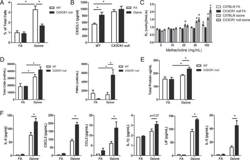
Ozone exposure in CX3CR1-null mice. C57BL/6 (WT) and CX3CR1GFP/GFP (CX3CR1-null) mice were exposed to filtered air or 2 ppm of ozone for 3 h and then underwent analysis 12–24 h after exposure. A, Flow cytometric analysis of Gr-1 Macs in WT (open box) and CX3CR1-null mice (closed box) as a percentage of total cells at 24 h after exposure to filtered air (FA) or ozone. B, CX3CL1 protein expression was analyzed by ELISA in BAL from WT and CX3CR1-null mice 24 h after filtered air (open box) or ozone (closed box) exposure. C, AHR after increasing doses of methacholine in WT and CX3CR1-null mice 24 h after filtered air or ozone exposure. D, Total cells and neutrophils (PMNs) from BAL cell count differentials in WT (open box) and CX3CR1-null mice (closed box) 24 h after filtered air or ozone exposure. E, Analysis of total protein in BAL from WT (open box) and CX3CR1-null mice (closed box) 24 h after filtered air or ozone exposure. F, Analysis of cytokines by multiplex from concentrated BAL fluid in WT (open box) and CX3CR1-null mice (closed box) 12 h after filtered air or ozone exposure. Data for AHR are from n = 7 WT FA, n = 7 CX3CR1-null FA, n = 12 WT O3, and n = 14 CX3CR1-null O3 (#p < 0.05 for WT or CX3CR1-null O3 versus FA control, *p < 0.05 for WT versus CX3CR1-null O3 exposed, &p < 0.05 for WT versus CX3CR1-null FA exposed). Data for other experiments are from three to eight mice per group (WT-FA, WT-ozone, CX3CR1-null–FA, and CX3CR1-null–ozone).
