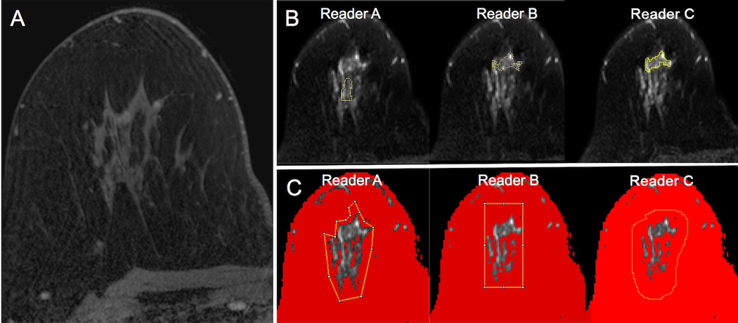
Figure 2.
An example of regions of interest (ROIs) placed on diffusion weighted images (b = 800 s/mm2) by three readers for a suspicious mass in the right breast as depicted on the subtracted axial post-contrast image (red box, A), biopsy-proven benign apocrine cystic metaplasia. All three readers placed ROIs (yellow lines) encompassing the lesion using both manual (B) and semi-automated (C) methods. Using the semi-automated thresholding tool, voxels of lower signal intensity were first masked out by each reader to be excluded from analysis (depicted in red) (C). Note the moderate variability in manual ROI size and shape between readers when attempting to include the lesion without including adjacent voxels of fat. By comparison, the voxels selected for analysis using the semi-automated method agreed well across readers.

