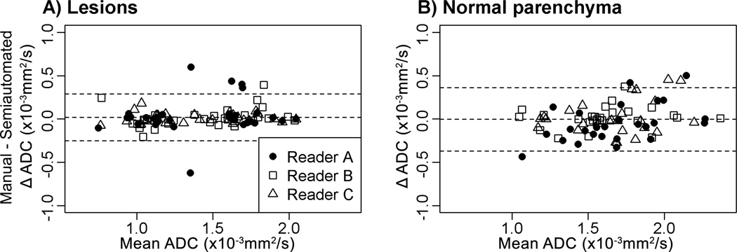
Figure 4.
Comparisons of ADC measures (×10−3 mm2/s) by manual and semi-automated methods for lesions (A) and normal tissue (B). For both lesions and normal tissue, the middle horizontal line (the average difference of readers’ ADC measures with manual and semi-automated methods) is close to zero, indicating that the use of the semi-automated method did not result in systematic bias.

