Figure 1.
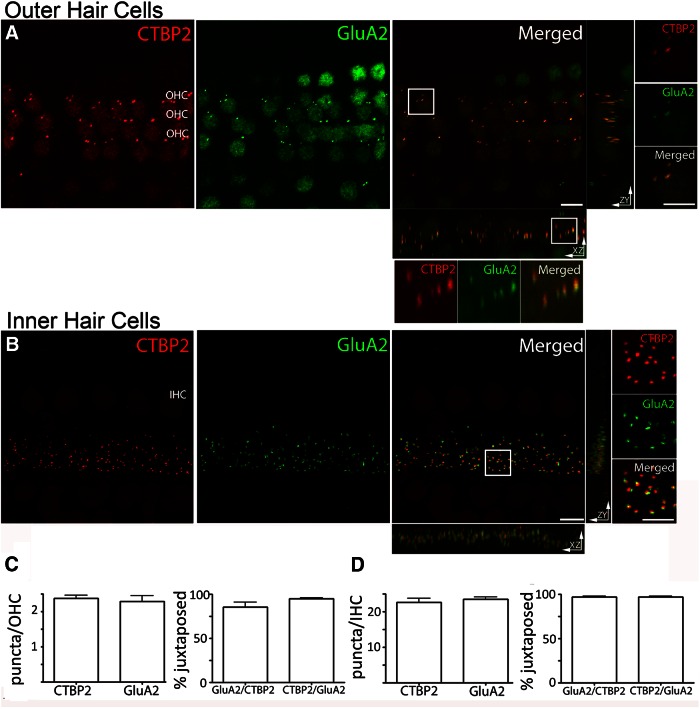
Ribbons and AMPAR clusters in cochlear whole mounts, and maximum intensity projections of confocal z-stacks of the medial region of the organ of Corti from an adult rat viewed from the endolymphatic surface including 24 adjacent OHCs and 5 IHCs. A, OHCs: immunolabel for the presynaptic ribbon marker (CtBP2, red channel). Immunolabel for the postsynaptic marker GluA2 (green channel). Merged and magnified inserts: CtBP2 and GluA2 puncta overlapped in the x- to y-plane. Rotation to the z- to x-planes or z- to y-planes reveals displacement between presynaptic and postsynaptic markers. B, IHCs: presynaptic and postsynaptic immunolabels. CtBP2 (red) and GluA2 (green) immunopuncta were consistently juxtaposed at the IHCs. Magnified insert in the x- to y-plane shows clear separation of presynaptic and postsynaptic labels. The x- to z-labels and z- to y-labels were not as well segregated as those in OHCs. C, Quantification of the number and the percentage of juxtaposed CTBP2 and GluA2 puncta in OHCs. D, Quantification of the number and the percentage of juxtaposed CtBP2 and GluA2 puncta in the IHCs. n = 3-9 independent preparations; 50 IHCs, 72 OHCs for A–D. There were no statistically significant differences in number or correlation among the immunolabels (one-way ANOVA test, p > 0.05). Scale bars: A, B, 5 µm; magnified inserts, 2.5 µm.

