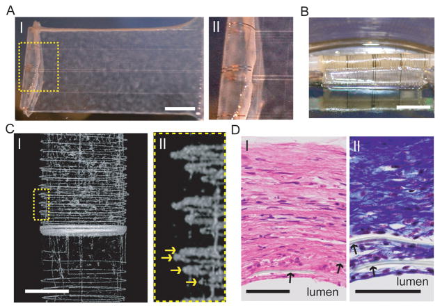
Figure 6. Nanoelectronics integrated into synthetic tissue.
(A) (I) Photograph of a single HASMC sheet cultured with sodium L-ascorbate on a nanoES. (II) Zoomed-in view of the dashed area in (I), showing metallic interconnects macroscopically integrated with cellular sheet. (B) Photograph of the vascular construct after rolling into a tube and maturation in a culture chamber for 3 weeks. (C) (I) Micro-computed tomograph of a tubular construct segment. (II) Zoomed-in view of (I). Yellow arrows mark the individual nanowire FET-containing layers of the rolled construct. Scale bar, 1 mm. (D) (I) Hematoxylin & eosin and (II) Masson Trichrome (;collagen is blue) stained sections of nanoelectronic-HASMC hybrid (~ 6 μm thick) cut perpendicular to the tube axis; lumen regions are labeled. Black arrows mark the positions of SU-8 ribbons of the nanoES. Scale bars, 50 μm.

