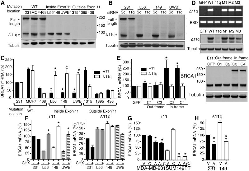
Figure 1. BRCA1 exon 11 mutant cell lines express BRCA1-Δ11q.
(A) Cell lines were analyzed for BRCA1 and tubulin levels by Western blot. *Predicted BRCA1 locations, molecular weights are indicated.
(B) Cells were treated with scrambled (Sc) or BRCA1-Δ11q (11q) siRNA and analyzed by Western blot.
(C) Exon 11 containing (+11) BRCA1 transcripts and the BRCA1-Δ11q (Δ11q) isoform were detected using qRT-PCR. Values were normalized to a HKG, expressed as a percentage of MDA-MB-231 cells.
(D) 293T cells were transfected with either GFP-control or BRCA1-minigene reporter constructs that were WT or carrying mutations that disrupted the cryptic 11q splice site (11q), or with frameshift mutations (M1:2288delT; M2:2529C>T; M3:3960C>T). BRCA1-Δ11q-reporter mRNA and protein expression was measured by RT-PCR (above) and Western blot (below), see Supplementary Fig. S3.
(E) CRISPR/Cas9 targeting the mutation-containing region of exon 11 (sg_exon11) generated SUM149PT clones (C) 1-4, see Supplementary Fig. S4. +11 and Δ11q mRNA was measured using qRT-PCR (left), values were normalized to a HKG and expressed as a percentage of sg_GFP control cells. BRCA1 protein was detected by Western blot (right).
(F) The indicated cell lines were treated with vehicle or 10 μg/ml CHX for 5 hours followed by assessment of +11 (left) and Δ11q (right) levels by qRT-PCR.
(G) Cells were treated with either vehicle (V), 10 μg/ml CHX (C), 5 μg/ml ACT (A) or A and C simultaneously for 5 hours and assessed for +11 expression by qRT-PCR.
(H) Cells were treated with either vehicle (V), or 5 μg/ml ACT (A) for 5-hours and assessed for Δ11q expression by qRT-PCR. *P < 0.05.

