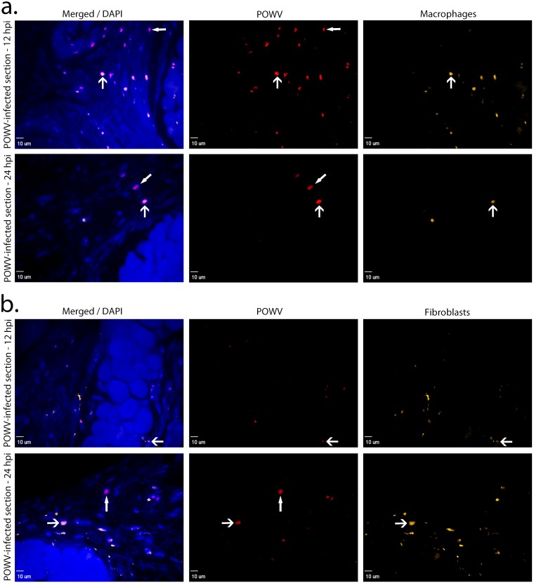
Fig 4. Immune cells detected at the POWV-infected Ixodes scapularis feeding sites, 12 and 24 hpi.
(A). Images of skin at the POWV-infected tick feeding site where macrophages are shown in orange and POWV-infected cells are shown in red. The F4/80 marker was used for macrophage detection. (B). Images of skin at the POWV-infected tick feeding site where fibroblasts are shown in orange and POWV-infected cells are shown in red. The vimentin marker was used for fibroblast detection. Scale bars represent 10 μm. DAPI (4’,6-diamidino-2-phenylindole) was used for nuclear counterstaining. Open arrowheads indicate POWV-infected macrophages or POWV-infected fibroblasts. Closed arrowheads indicate immune cells that are POWV-infected but not macrophages or fibroblasts.

