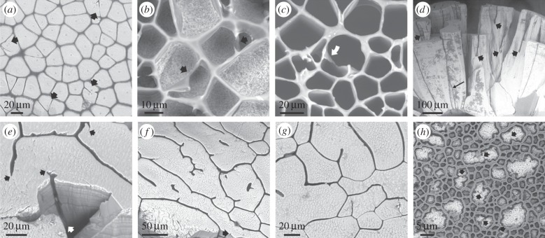
Figure 4.
Instances of advancing membranes in Pinctada margaritifera (a–c,h) and pearls of Pinna (d–g). (a) General aspect of the external shell surface, with the periostracum removed. Some prismatic units contain two or three crystals (arrows), which are partly fused. (b,c) Instances in which incomplete membranes meet and fuse during shell growth, thus delineating two different prismatic units. In (c), one of the incomplete membranes has rotated and stuck to the cell wall (arrow). (d) Radial fracture through a pearl of Pinna. During growth, some units wedge out (long thin arrow), but the pattern is dominated by the splitting of prismatic units owing to the initiation of organic membranes (thick arrows). (e) Oblique view of the fracture and surface of the same pearl as in (d). The membrane seen on the fracture surface initiates at the position of the white arrow. The tips of the membranes and of their minor branches consistently coincide with boundaries between crystalline domains (black arrows). (f,g) Two views of the growth surface of another pearl. As in (e), the tip of every membrane branch always continues into a nanocrystalline domain boundary. Isolated membranes appear within the cells interior in (f). The arrow in (f) points to the position of a very incipient subsidiary membrane. (h) Locally developed meshwork, possibly delineating individual crystalline domains (arrows point to some crystal boundaries). Panels (a–c) are views of the external shell surfaces, and (e–h) are views of the growth surfaces.

