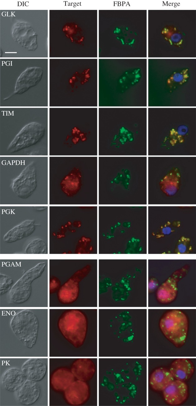
Figure 2.

Cellular localization of glycolytic enzymes in Diplonema papillatum using immunofluorescence microscopy. Cells were labelled with polyclonal antisera to GLK, PGI, TIM, GAPDH, PGK, PGAM, ENO or PK (Target, red) and with antisera to D. papillatum FBPA as a peroxisomal marker (FBPA, green), and the reactions were visualized using Alexa Fluor 568- and 488-conjugates, respectively. Images of the same cells were merged together with the labels of Hoechst 33342 (blue) to visualize the nucleus (Merge). DIC, differential interference contrast image; Merge, merged image. Scale bar, 10 µm.
