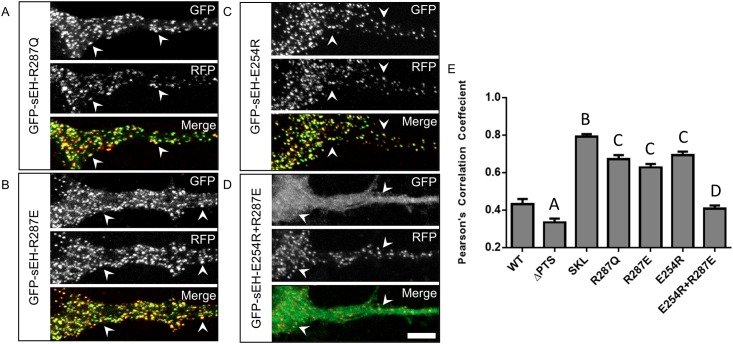
Fig 3. Disrupting dimerization preferentially localizes sEH to peroxisomes while rescuing dimerization restores wildtype sub-cellular distribution.
Primary neurons co-transfected with GFP-sEH fusion protein and peroxisome marker RFP-SKL. (A) GFP, RFP and merged image from a neuron transfected with GFP-sEH-R287Q. GFP-sEH-R287Q fusion results in a punctate pattern that highly co-localizes with RFP-SKL (white arrowheads) indicative of peroxisome localization. (B) GFP, RFP and merged image from a neuron transfected with GFP-sEH-R287E. GFP-sEH-R287E fusion results in a punctate pattern that correlates with RFP-SKL (white arrowheads) indicative of peroxisomal localization. (C) GFP, RFP and merged image from a neuron transfected with GFP-sEH-E254R. GFP-sEH-E254R fusion results in a punctate pattern that co-localizes with RFP-SKL (white arrowheads), indicative of peroxisomal localization. (D) GFP, RFP and merged image from a neuron transfected with GFP-sEH-E254R+R287E. GFP-sEH-E254R+R287E fusion results in a dual diffuse and punctate staining pattern. GFP-sEH-E254R+R287E puncta colocalize with RFP-SKL (white arrowheads) reflecting the dual distribution of GFP-sEH-E254R+R287E between the cytosol and peroxisomes similar to GFP-sEH-WT. (E) Quantification of peroxisome localization for each GFP-sEH construct with Pearson’s correlation coefficient. A indicates p<.05 compared with WT, SKL, R287E, and E254R. B indicates p<.05 compared with WT, ΔPTS, R287Q, R287E, and E254R and E254R+R287E. C indicates p<.05 compared with WT, ΔPTS, and E254R+R287E. D indicates p<.05 compared with ΔPTS, SKL, R287Q, R287E, and E254R. Scalebar is 5μm.

