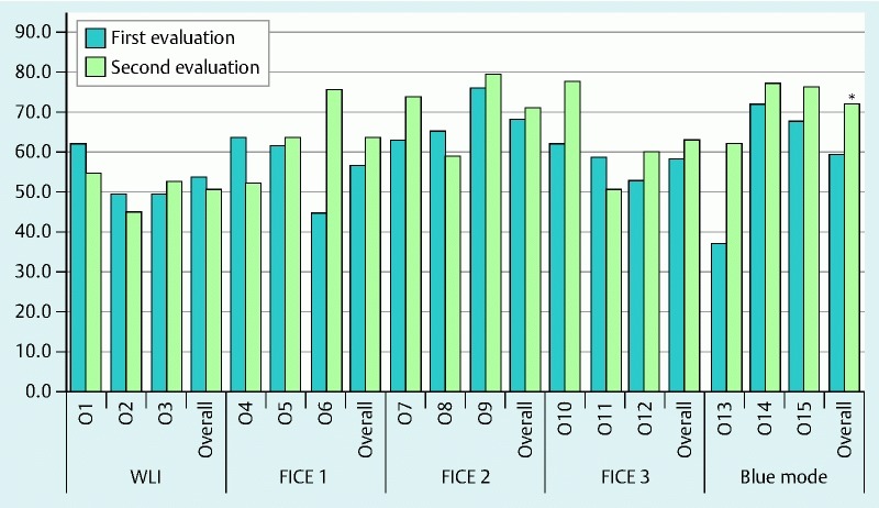
Fig. 3.

Comparison of accuracy in correctly identifying the ulcerative images for all readers at their first and second evaluations. The investigators were randomized for the second reading to evaluate the study images in WLI (O1 – 3), FICE 1 (O4 – 6), FICE 2 (O7 – 9), FICE 3 (O10 – 12) and Blue mode (O13 – 15). Of the overall evaluations, statistical significance was obtained only for Blue mode (*); WLI, white light imaging; O, observer.
