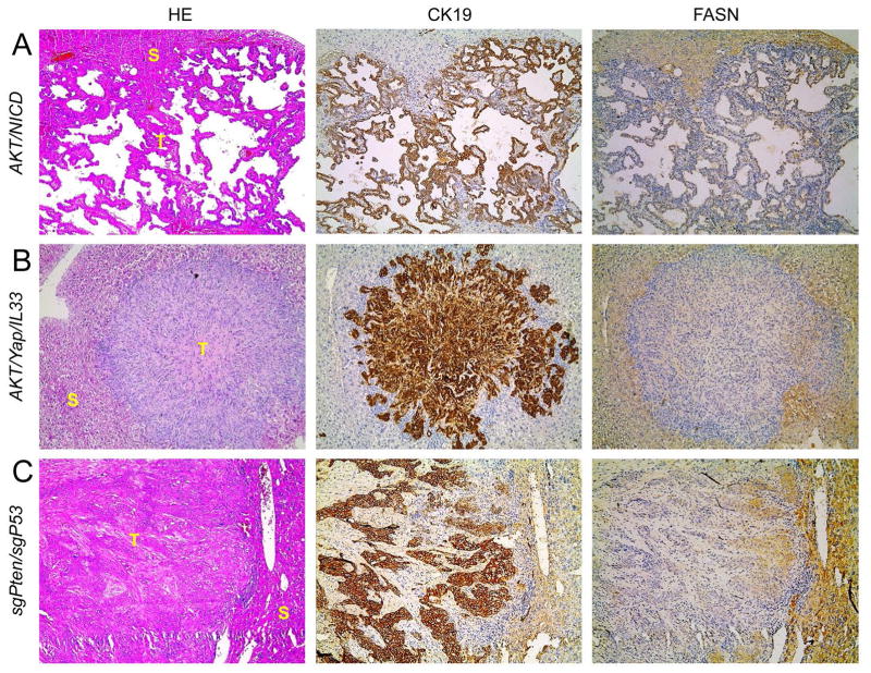
Figure 2.
FASN expression pattern in AKT/NICD, AKT/Yap/IL33, and sgPten/sgP53 mouse models, as detected by immunohistochemistry. Similar to human ICC specimens, immunoreactivity for FASN is either faint or absent in ICC (T), whereas it is preserved in non-tumorous surrounding liver (S). Original magnification: 100X. Abbreviation: HE, hematoxylin and eosin staining. At least 3 mice in each tumor group were analyzed by immunostaining.

