
Figure 4. Reduction in CCl4-induced liver fibrosis in miR-155 KO mice.
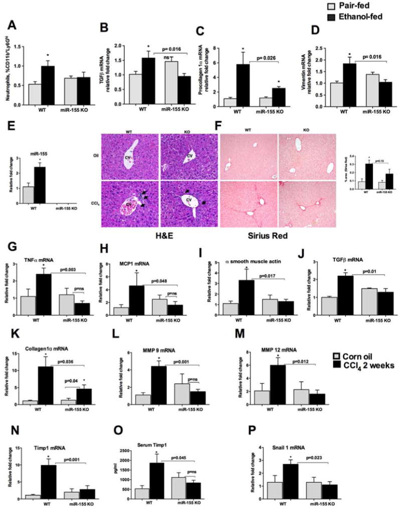
WT or miR-155 KO mice were fed with Lieber DeCarli diet. MNCs were stained for neutrophils (A). Liver RNA was used to determine TGFβ (B) procollagen 1α (C) and vimentin (D) mRNA levels by qPCR. 18s was used to normalize Cq values. Data is presented as mean ± SEM. * indicates p<0.05 vs. pair fed mice, ns: non significant. WT or miR-155 KO mice (n=6) were treated either with corn oil or CCl4 for 2 weeks as described in methods. Total RNA was used to determine miR-155 levels by qPCR (E). Formalin fixed liver sections were stained with Hematoxylin and eosin (F, left panel) and Sirius red stain (F, right panel). Slides were observed under light microscope (100X) and representative slides are shown. RNA was used to determine TNFα (G), MCP1 (H), α smooth muscle actin (I), TGF β (J), collagen 1α (K), MMP9 (L), MMP12 (M), Timp1 (N), Snail1 (P) mRNA levels by qPCR. Timp1 levels were measured from plasma by ELISA (O). 18s or SnoRNA202 was used to normalize Cq values for mRNA, and mouse miR-155 respectively. Data is presented as mean ± SEM. * indicates p<0.05 vs. corn oil treated mice.