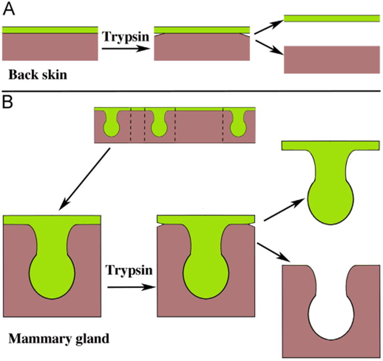
Fig. 4.

Schematics of 14-day embryonic back skin (A) and 13-day embryonic mammary glands (B). The flat planar epidermis shows evidence of “loosening” following trypsinization so that clean separation of epithelium and mesenchyme can be achieved (A). A strip of skin with three mammary epithelial buds is shown and in turn trimmed to size (B). Following trypsinization, the edges of the epidermis are separating, which indicates that the tryptic digestion was successful, thus allowing clean separation of the epithelium and mesenchyme. Mesenchyme = brown, epithelium = green. (Adapted from (Cunha, 2013b) with permission). (For interpretation of the references to color in this figure legend, the reader is referred to the web version of this article.)
