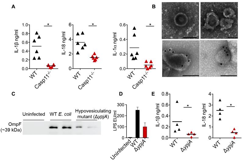
Figure 7. OMV are Essential for the Activation of Cytosolic LPS Sensing in vivo.
(A) IL-1β, IL-18, and IL-1α levels in the plasma of wild-type and caspase-11−/− mice injected i.p. with 100 μg of OMV. Mice were first primed with 200 μg poly(I:C) for 6 h (i.p.). Cytokine levels were assessed 6 h post OMV injection (n=6).
(B) Negative staining (top row) and LPS immunogold (bottom row) TEM for OMV in the peritoneal lavages from wild-type mice infected i.p with 109 CFU of E. coli for 12 h (images were from two different mice). Scale bar = 0.1 μM.
(C-D) Immunoblot for OmpF (C) and LAL assay for LPS (D) in OMV isolated from the peritoneal lavages of mice infected with 109 CFU of wild-type or ΔypjA strain of E. coli for 12 h. Each lane represents one mouse.
(E) IL-1β and IL-18 levels in the plasma of wild-type mice infected with 109 CFU of wild-type or ΔypjA strain of E. coli for 12 h (n=4).
Data are presented as mean±SEM of one experiment representative of two experiments. See also Figure S6.

