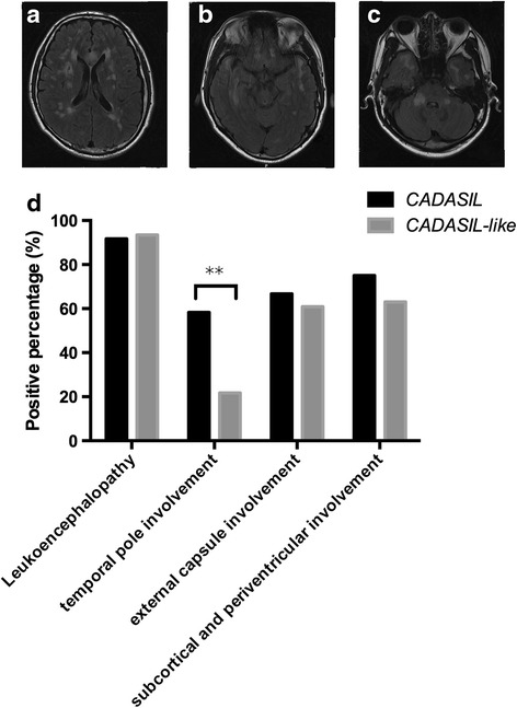
Fig. 3.

T2-Flair magnetic resonance images from the same 42-year-old patient with NOTCH3 p.Arg153Cys showing diffuse white matter hyperintensities in (a) bilateral centrum semiovale, (b) temporal pole and (c) pedunculus cerebellaris medius. (d) indicated the percentage of the positions involved (**p < 0.01)
