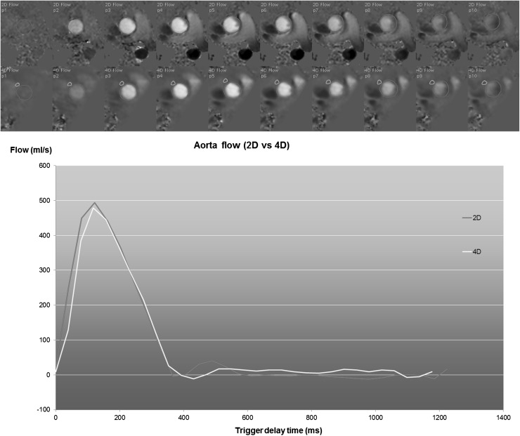
Fig. 5.
Example of aortic flow curves of a healthy subject derived from conventional 2D phase-contrast MRI (2D Flow) and from a reformatted 4D Flow acquisition. The 4D Flow acquisition was reformatted into a through-plane encoded view, identical to the corresponding 2D Flow acquisition. The top two rows depict the systolic through-plane velocity-encoded images (the first 10 frames out of 30) for 2D Flow and 4D Flow MRI, respectively. Contours are defined around the aortic lumen (shown in red). In the 4D Flow-derived images, an additional contour is defined in a region of tissue adjacent to the aorta for velocity offset correction. A good agreement is observed between the two flow curves. The stroke volume derived from 2D Flow is 105 ml and from 4D Flow is 103 ml (Color figure online)

