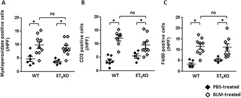
Fig. 4.

Infiltration of inflammatory cells in the dermis of WT and ETBKO mice after BLM treatment. The average cell counts of a myeloperoxidase, b CD3, and c F4/80-positive cells in the dermis. The cells were counted per field of view at 100× magnification; n = 5–10 mice per group. (* p < 0.05). BLM bleomycin, ET B KO endothelin type B receptor knockout, HPF high-power field, PBS phosphate-buffered saline, WT wild-type
