Fig. 1.

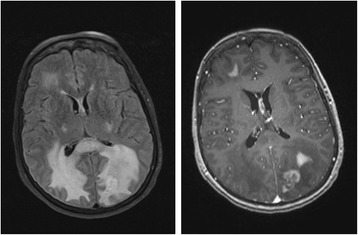
Brain MRI (October 2009): axial FLAIR (left) and axial T1 post contrast (right) revealing right frontal and left parieto-occipital enhancing lesions with surrounding edema

Brain MRI (October 2009): axial FLAIR (left) and axial T1 post contrast (right) revealing right frontal and left parieto-occipital enhancing lesions with surrounding edema