Fig. 3.

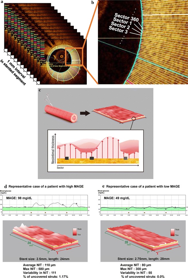
Three-dimensional assessment of the neointimal distribution. a The lumen and stent areas were manually traced every 1 mm. b The neointimal area was divided into 360 circumferential sectors (each section is 1°, which is referenced from the lumen’s center). The mean neointimal thickness (NIT) was calculated for each sector in each 1-mm section, and the same sector’s thicknesses were compared throughout the entire stented segment. c To visualize the two-dimensional layout, the stent surface was cut along the longitudinal direction and flattened into a rectangular shape. The roughness of the neointima (variability in NIT) within the whole stent was evaluated based on the standard deviation (SD) of the NITs from the same 1°-sector along the entire stented segment. Representative cases are shown for a patient with high mean amplitude of glycemic excursion (MAGE) (d) and a patient with low MAGE (e)
