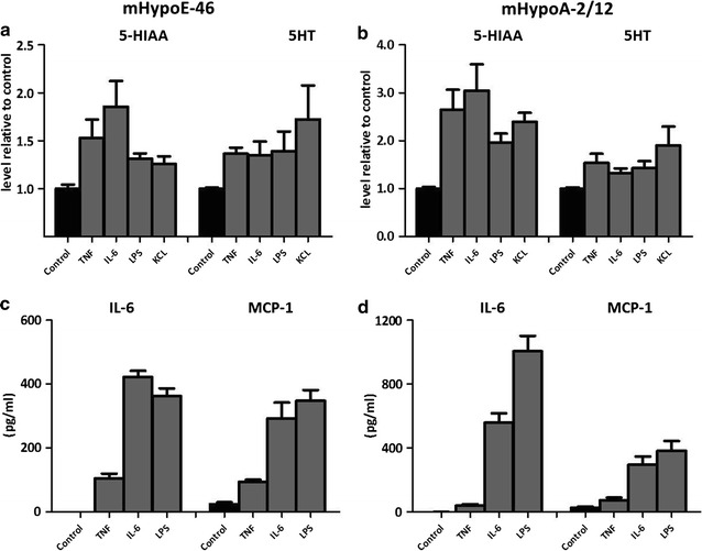
Fig. 2.

Effect of TNFα, IL-6 and LPS on 5-HIAA, 5HT, IL-6 and MCP-1 in HypoE-46 and mHypoA-2/12 cells. Murine derived hypothalamic cell lines were 24 h exposed to various concentrations TNFα (100 pg/ml), IL-6 (100 pg/ml) and LPS (1 μg/ml). KCL was used to depolarize cells (positive control for 5HT). a, b Intracellular 5-HIAA and secretion of 5HT in HypoE-46 (a) and HypoA-2/12 cells (b). c, d Production of IL-6 and MCP-1 in HypoE-46 (c) and HypoA-2/12 cells (d). All treatments were significantly different from untreated controls. Data are expressed as mean ± SEM (n = 3)
