Fig. 1.

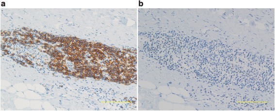
Immunohistochemical staining showing (a) numerous CD20-positive B cells and (b) scant CD3-positive T cell infiltration in periprosthetic tissue. Scale bar = 100 μm

Immunohistochemical staining showing (a) numerous CD20-positive B cells and (b) scant CD3-positive T cell infiltration in periprosthetic tissue. Scale bar = 100 μm