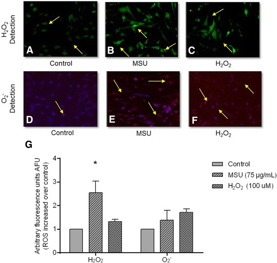
Fig. 2.

Monosodium urate (MSU) crystals increase reactive oxygen species (ROS) in synoviocytes. Arrows indicate intracellular H2O2 formation, which is revealed by DCFH oxidation (green fluorescence) in untreated fibroblast-like synoviocytes (FLS) (a); FLS treated with MSU crystals at 24 h (b), and FLS treated with H2O2 at 30 minutes (c). Arrows indicate O2 - intracellular production by oxidation of dihydroethidium (DHE) (red fluorescence) in untreated FLS (d); FLS treated with MSU crystals (e), and FLS treated with H2O2 (f). Bars show quantification of DCFH and DHE fluorescence: data are reported as units of arbitrary fluorescence (UAF) (g). Values are expressed as the mean ± standard deviation; *P < 0.05 vs control
