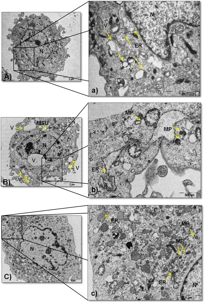
Fig. 5.

Ultrastructural changes in synoviocytes. A Ultrastructure of an untreated fibroblast-like synoviocytes (FLS). a Magnified view of the section is indicated by a black box showing the nucleus (N), endoplasmic reticulum (ER) and vacuoles (V) highlighted with arrows. B FLS treated with monosodium urate (MSU) crystals at 75 μg/mL exhibiting N, swollen vesicular structures of different sizes, and MSU crystal cavity. b A high-magnification image showing misfolded proteins (MP) aggregates and ER indicated with arrows. C FLS treated with H2O2 at 100 μM showing N. c A magnified view of the section is indicated by a black box showing MP aggregates, ER and N. Results are representative of one of five separate experiments with FLS from different patients
