
Fig. 9.
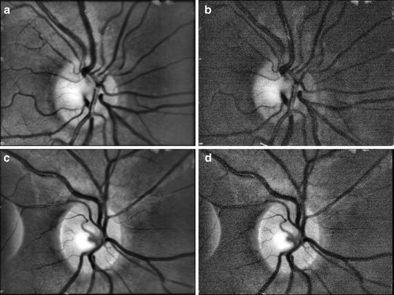
Two examples of averaged retinal images from two sequences (a, c) and single frame from corresponding sequence (b, d). The CLAHE method has been applied on each image to increase the contrast
Official websites use .gov
A
.gov website belongs to an official
government organization in the United States.
Secure .gov websites use HTTPS
A lock (
) or https:// means you've safely
connected to the .gov website. Share sensitive
information only on official, secure websites.
Two examples of averaged retinal images from two sequences (a, c) and single frame from corresponding sequence (b, d). The CLAHE method has been applied on each image to increase the contrast