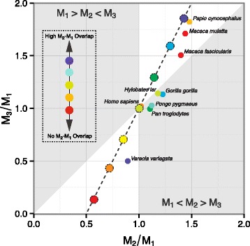
Fig. 5.

Developmental timing of lower molar calcification in primates (n=9). Points are species mean molar proportions. Colors denote degree of temporal overlap in M 2– M 1 calcification start times. Dashed line indicates DIC model’s predicted relationship between molar proportion ratios: M 3/M 1=2(M 2/M 1)−1. White regions are locations in molar proportion morphospace consistent with the DIC model: a high a/i region where M 1<M 2<M 3 and a low a/i region where M 1>M 2>M 3
