
Figure 2.
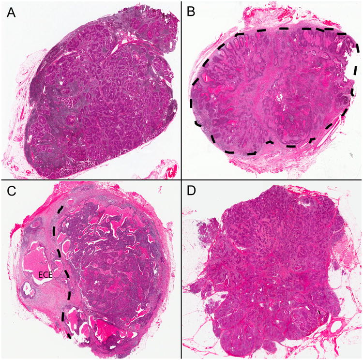
Histopathologic examples of extracapsular extension Grades from the defined classification system. A) Tumor cells limited to lymph node parenchyma without alteration of the capsule or nodal architecture (Grade 0). B) Tumor cells limited to lymph node but with expansion of the node and development of a thickened capsule/pseudocapsule (designated by dashed line) around them (Grade 0c). C) Tumor cells invading into perinodal soft tissue (beyond dashed line and indicated by “ECE”) but with at least partial preservation of the lymph node (Grade 1). D) Tumor cells growing as irregular collections in the neck soft tissues without any histologic evidence of residual nodal parenchyma (Grade 2 or “soft tissue metastasis”) (all images hematoxylin and eosin stained; 15× magnification).
ECE = extracapsular extension