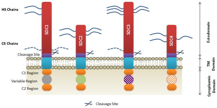
Figure 2. The core-domain structure of syndecans.
The extracellular domain is depicted in red and both heparan and chondroitin sulphate chains are indicated by the blue lines. Cleavage sites are indicated by purple blocks, followed by a short transmembrane domain in blue. Each syndecan has a short cytoplasmic domain in which a variable region is flanked by two highly conserved regions termed C1 and C2. Cleavage of the ectodomain can occur via numerous metalloproteinases such as MMP2, 3, 7 and 9, as well as proteases thrombin and plasmin. HS, Heparan sulphate; CS, Chondroitin sulphate; SDC, syndecan; TM, transmembrane.

