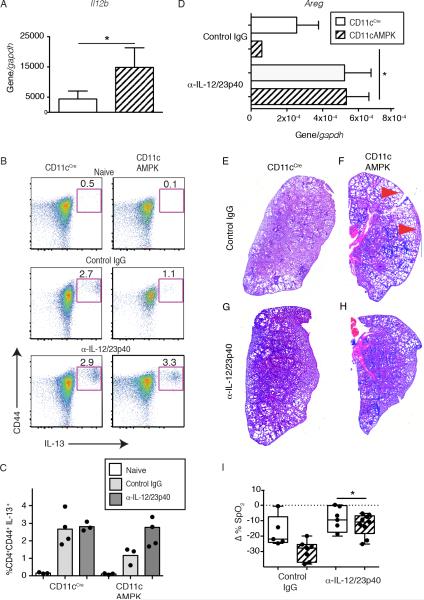
Fig. 6. Neutralization of IL-12/23 p40 in CD11cAMPK mice restores Type 2 responses and lung repair.
A) Comparison of whole tissue mRNA transcript levels for Il12b from sorted pulmonary CD11c+ cells between CD11cCre and CD11cAMPK mice at 9 dpi with N.b. B) Representative dot plots showing the percentage of CD4+CD62LoCD44HiIL-13+ cells within the spleen of naïve (top) or at 9 dpi following IgG2a isotype mAb (middle), or anti-IL-12/23p40 mAb-treatment (bottom) mice. C) Dot plot showing the percentage of CD4+CD62LoCD44HiIL-13+ cells within the spleen of naïve (white), or at 9 Dpi following isotype mAb (gray), or anti-IL-12/23p40 mAb-treatment (charcoal) mice. D) Areg mRNA transcript of lung tissue from CD11ccre (solid) and CD11cAMPK (hatched) mice 9 dpi with N. b. L3. Graph indicates relative expression over the Gapdh reference gene. H&E-stained cross-section of left lobe of lung (200x magnification) from G, H) isotype mAb or I, J) anti-IL-12/23p40 mAb-treatment. Red arrows indicate areas of lung injury. F) Change in % SpO2 at 8 dpi following treatment with 3 doses of 1mg anti-IL-12/23p40 mAb or IgG2a isotype mAb. Data represent two independent experiments containing 3-5 mice per group. *p<0.05, **p<0.01

