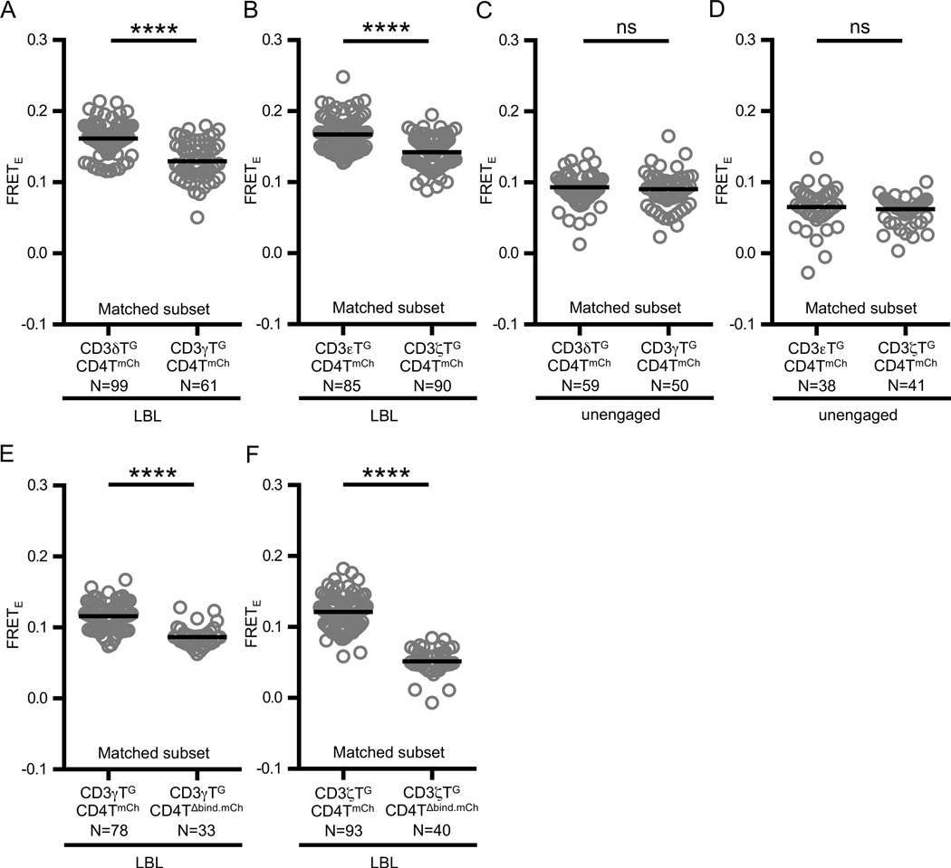
Figure 3. CD4 adopts an ordered arrangement with respect to the CD3 subunits.
(A–B) Agonist pMHC engagement results in differential FRETE between CD4 and the CD3 subunits. Experimental M12 cells were adhered to supported lipid bilayers presenting MCC:I-Ek and ICAM-I (LBL) as labeled and described in the text.
(C–D) Differences in FRETE depend on ligand engagement. Experimental M12 cells were adhered to glass coverslips (unengaged).
(E–F) FRET between CD4 and (E) CD3γ or (F) CD3ζ subunits is dependent on CD4 D1 domain-MHC interactions. Experimental M12 cells were adhered to supported lipid bilayers presenting MCC:I-Ek and ICAM-I (LBL) as labeled and described in the text.
Circles represent individual cells, N= number of cells analyzed, and black bars represent median values. Data are representative of at least two experiments per condition. Subset analysis and subset criteria are as detailed in Materials and Methods (****p<0.0001; Mann-Whitney).

