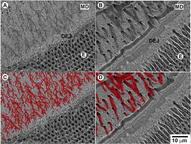
Fig. 6.
Dspp–/– teeth present with major defects at the DEJ, mantle dentin and inner enamel. BS SEM images of etched erupted portions of incisors mice, polished in the transverse plane and resin cast, from one-month-old mice. A. Dspp+/+ and B. Dspp–/–. C and D correspond to images A and B with the odontoblasts processes colored for a better visual perception. All micrographs are taken at the same magnification. E—enamel, MD—mantle dentin. (For interpretation of the references to color in this figure legend, the reader is referred to the web version of this article.)

