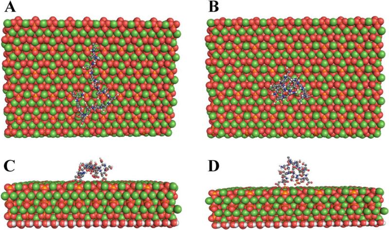
Figure 3.
Molecular Dynamics Simulation: The most commonly repeated peptide of Dentin phosphophoryn (P5 Ace-SSDSSDSSDSSDSSD-NH2 and P5P Ace-SSDpSpSDpSpSDpSpSDpSpSD -NH2) binds in a more extended form to the 100 surface of hydroxyapatite when phosphorylated (A,C) than when unphosphorylated (B,D). A and B show the mineral surface, B and D are viewed perpendicular to the surface. In the mineral model red circles are oxygen (O) atoms, orange circles phosphorus (P) atoms, white circles hydrogen (H) atoms and green circles calcium (Ca) atoms. Water is not shown but was included in the model. For details of the modeling procedures see Villarreal-Ramirez et al., 2014 [108]).

