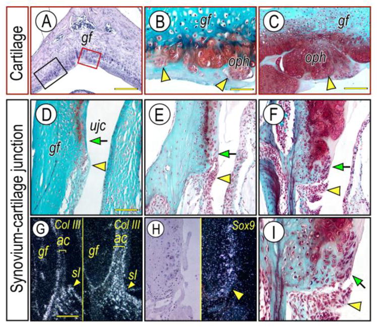
Figure 3. Major steps of osteophyte development near articulating cartilage and synovium-articular cartilage junction in Prg4−/− glenoid fossa.

Frontal sections prepared from TMJs of 3 month- (D; G, right panel), 6-month- (A, B, E), 10 month- (C, F, I) old Prg4−/− mice were stained with Hematoxylin & Eosin (A) and Safranin-O (B–F, I). Osteophyte development was investigated in red box area (articular cartilage) or black box area (synovium-cartilage junction) of the glenoid fossa (A) representing the articulating cartilage and synovium-articular cartilage junction, respectively. Note that multiple osteophyte-like chondrocyte clusters start forming in the articulating cartilage surface (B, arrowheads), and grow into osteophytes (oph) (C, arrowhead). Synovial cell mass (D–F; arrowhead) present at the synovium-articular cartilage junction before chondrocyte differentiation indicated by lack of Safranin-O staining and its integration into the developing osteophytes (F, I, arrow) in the advanced OA condition. Synovium-articular cartilage junction in F was magnified to reveal the synovial cell mass (I, arrowhead) and integration of synovial cells into developing cartilage/osteophyte (I, arrow). In situ hybridization with isotope-labeled RNA probe of type III collagen (Col-III) (G, left panel, control; right panel, mutant) and Sox9 (H). Note that Col-III transcripts are abundant in the synovial membrane and are much less at the articular cartilage surface, while a numerous number of Col-III-expressing cells are detectable in the Prg4−/− glenoid fossa. Sox-9 expressing chondroprogenitor cells detected in the synovium-articular cartilage junction in Prg4−/− glenoid fossa. Scale bars: 150 μm in A; 45 μm in B for B, I; 80 μm in C for C–F; 125 μm in G for G–H. gf, glenoid fossa; ujc, upper joint cavity; oph, osteophyte; sl, synovial lining cell; ac, articular cartilage.
