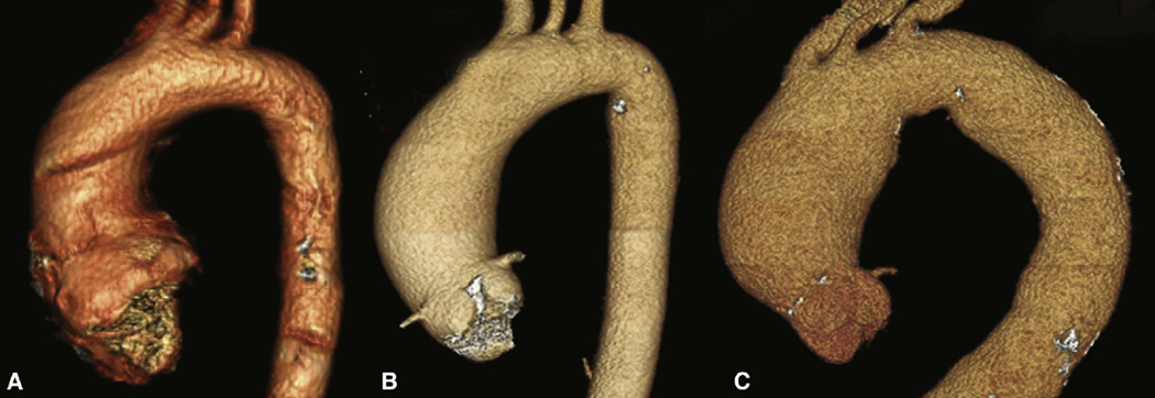
FIGURE 4.
Three-dimensional renderings of preoperative computed tomographic angiograms from A, a patient with type 0 bicuspid aortic valve, B, a patient with type 1 bicuspid aortic valve with right/left morphotype, and C, a patient with tricuspid aortic valve. The dilation patterns of all 3 patients with the type 1 right/left morphotype had the same pattern as depicted in B.

