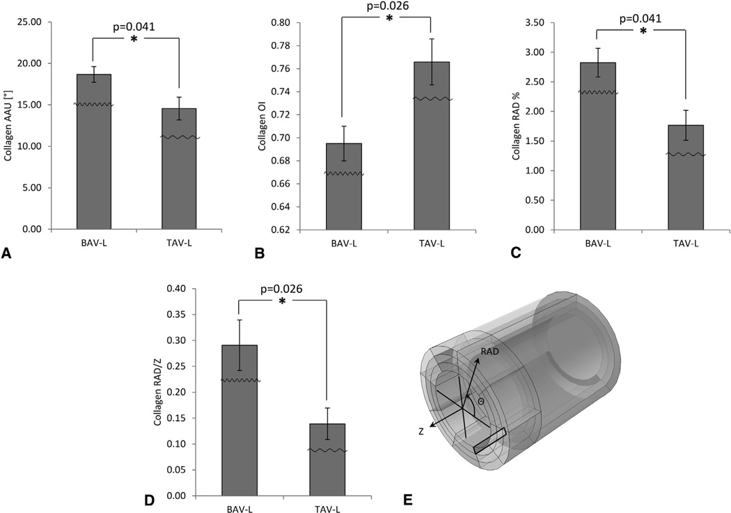
FIGURE 6.
Results of Mann-Whitney U tests (only the significant differences [P < .05] are presented) for the comparison of collagen microarchitecture between the 2 ascending thoracic aortic aneurysm phenotypes in the media-intima (med-int) layer in the longitudinal (Z) radial (RAD) plane of region L based on the output of the custom MATLAB written automated image-based analysis tool. A Amplitude of angular undulation of collagen (same trend also observed for collagen amplitude of angular undulation between bicuspid aortic valve (BAV) and tricuspid aortic valve (TAV) in the adventitia-media (adv-med) layer of Z-RAD and circumferential (Θ) RAD planes of region L; see first row of Table 2 for P = .026 in both cases; bar graphs not shown here), B Orientation index (OI) of collagen fibers, C, percent of collagen fibers oriented in the RAD-direction (same trend also observed for percent of elastin fibers oriented in the RAD-direction between BAV and TAV [P = .041] in the same layer [med-int], plane [Z-RAD], and region [L]; bar graphs not shown here), and D ratio of collagen fibers oriented in the RAD over Z-direction. The little waveforms overlaid onto the bars indicate how the waviness of the fibers is affected. E, The black-colored rectangle within the model tube indicates the layer (med-int), region (L), and plane (Z-RAD) across the thickness of the wall. Results are presented as mean ± standard deviation. *Significant difference. AAU, Amplitude of angular undulation; OI, orientation index; BAV-L, bicuspid aortic valve circumferential region with respect to left coronary sinus; TAV-L, tricuspid aortic valve circumferential region with respect to left coronary sinus.

