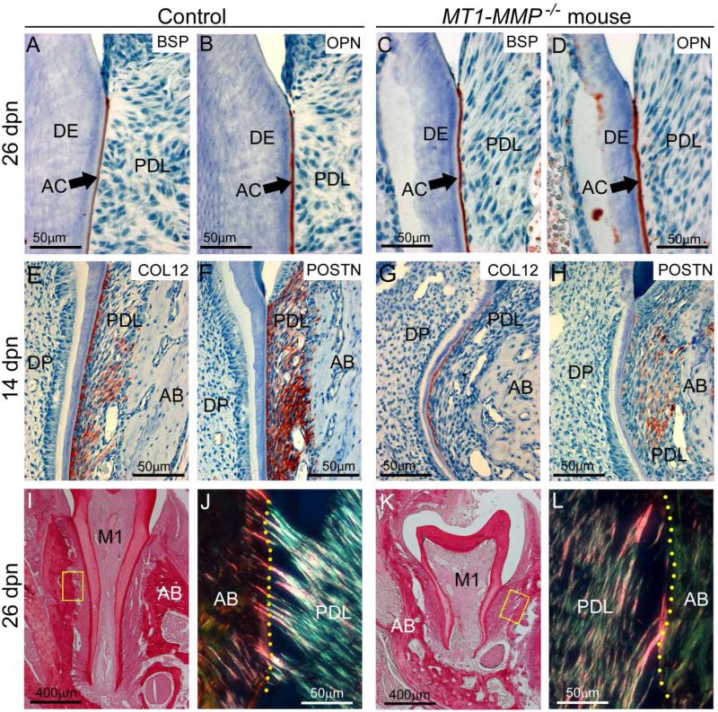
Figure 5. Loss of MT1-MMP disrupts periodontal structure.
Immunostaining and selective staining approaches were used to analyze periodontal development in WT control and MT1-MMP−/− mice. (A-D) BSP and OPN immunostaining indicate the presence the acellular cementum (AC) layer on the molar root surface in controls and MT1-MMP−/− mice. The PDL begins to organize in WT by 14 dpn, as shown by increased localization of ECM markers (E) collagen type XII (COL12) and (F) periostin (POSTN), whereas the MT1-MMP−/− mouse PDL shows little localization of (G) COL12 or (H) POSTN, though strong COL12 staining near the root surface reflects collagen fiber insertion into cementum. Picrosirius red staining (under non-polarized light) indicates the presence of fibrillar collagen in 26 dpn (I) WT and (K) MT1-MMP−/− first molars (M1), PDL, and alveolar bone (AB). Under polarized light, (J) WT PDL shows organized collagen fibrils in the PDL, and numerous insertions of Sharpey's fibers into the alveolar bone (yellow dotted line indicates the PDL-bone border). (K) MT1-MMP−/− PDL shows less organization, with loose collagen fibers running mostly parallel to the root surface, and few Sharpey's fibers that are also poorly embedded.

