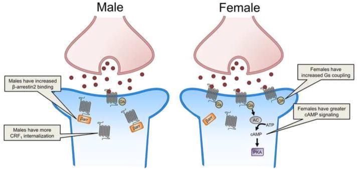
Figure 3.
This schematic depicts sex differences in the CRF1 receptor. The image on the left depicts increased β-arrestin2 binding to the CRF1 receptor (gray) and internalization in a male LC neuron (blue). The image on the right depicts CRF (red circle) binding to the CRF1 receptor (gray), which increases Gs coupling and signaling through the cAMP pathway in a female LC neuron (blue). AC, adenylyl cyclase; ATP, adenosine triphosphate; βarr., β-arrestin2; cAMP, cyclic adenosine monophosphate; PKA, protein kinase A

