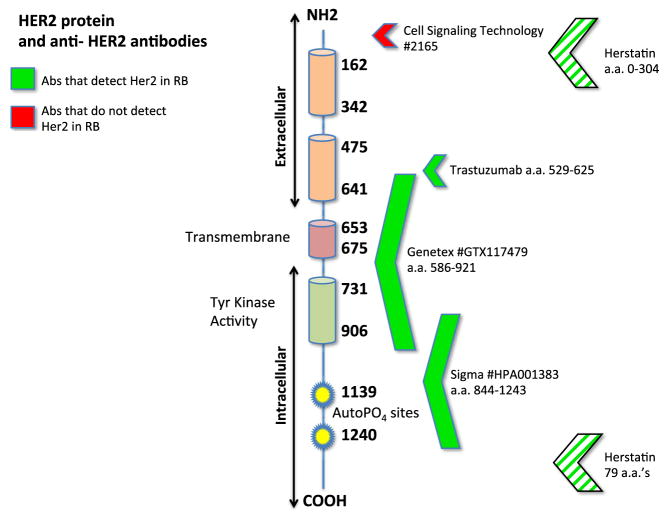
Fig. 8.
HER2 antibodies and the HER2 protein. Antibodies used for this study and their binding sites along the HER2 protein are shown. Green arrows indicate positive detection of HER2, while red arrows show negative results. Based on these results, it appears that the HER2 truncation is at the amino terminal end. Note that the Herstatin antibody recognizes a splice variant that includes both amino and carboxy-terminal ends (striped arrows)

