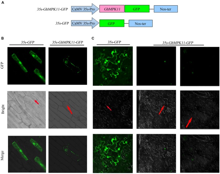
FIGURE 2.
The subcellular localization of GhMPK11 protein transiently expressed in onion epidermal cells and tobacco cells. (A) Schematic diagram of the 35S-GhMPK11-GFP fusion construct and the control 35S-GFP construct. GFP was fused to the C terminus of GhMPK11. Transient expression of the 35S-GhMPK11-GFP and 35S-GFP constructs in onion epidermal cells (B) and tobacco cells (C). Red arrows indicate the location of the nucleus. Green fluorescence was observed using a confocal microscope. Bar = 200 mm.

