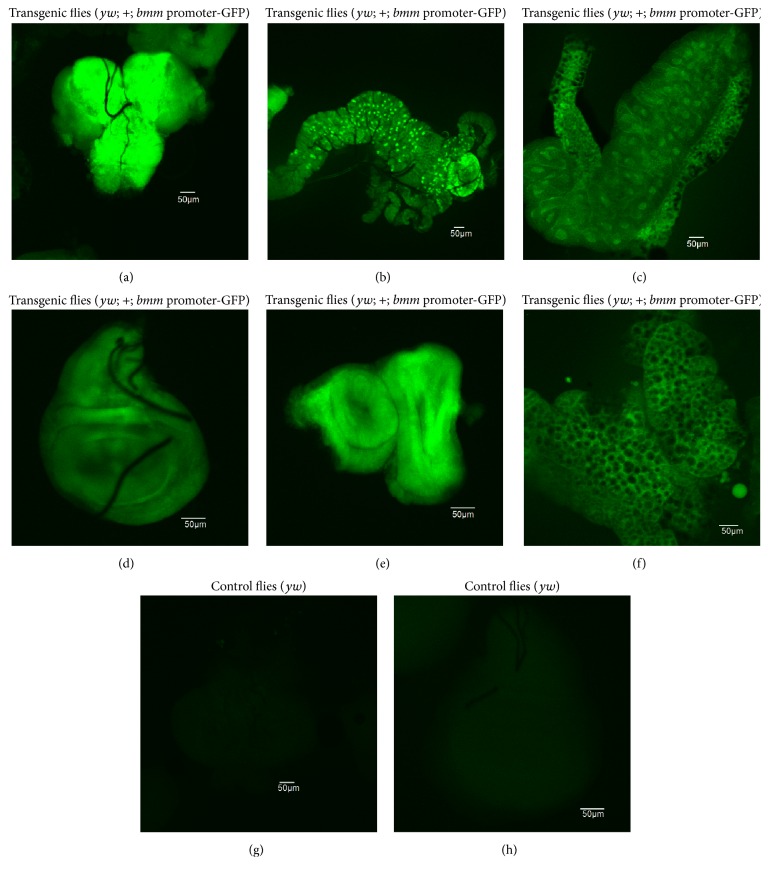
Figure 2.
The bmm promoter-driven expression of GFP in transgenic Drosophila (yw; +; bmm promoter-GFP). The various tissues of the third-instar larvae of transgenic flies were observed by fluorescence microscopy. The sections of brain lobe (a), gut (b), salivary gland (c), wing disc (d), eye disc (e), and lipid tissue (f) showed GFP signals. In contrast, the control fly (yw) showed no detectable GFP signal ((g) brain lobe; (h) wing disc).

