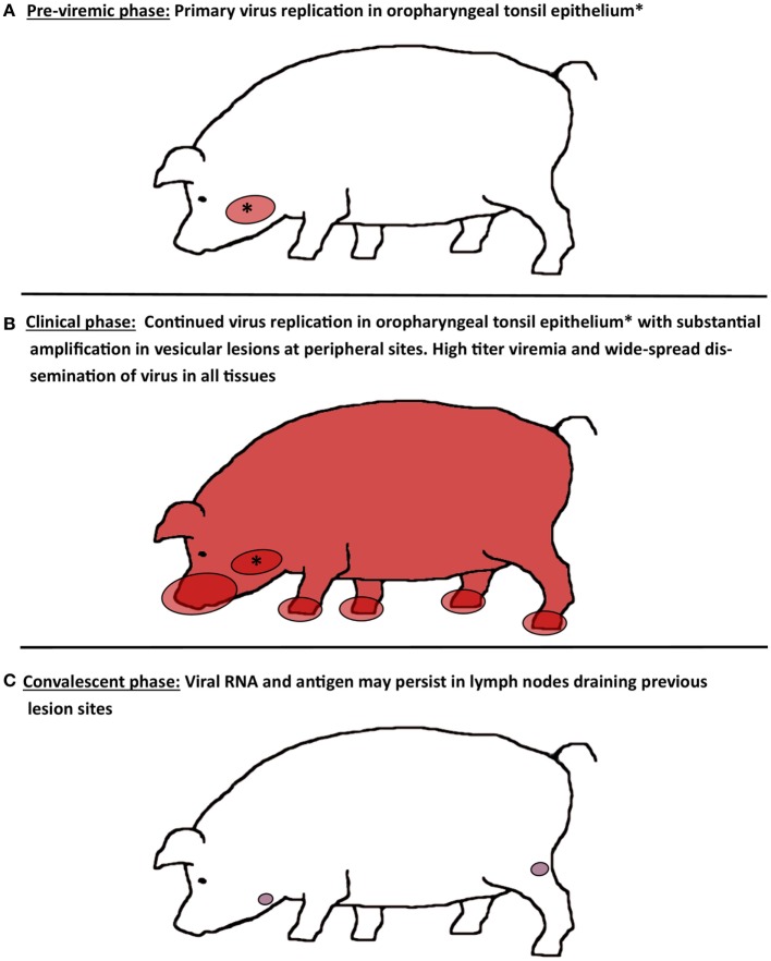
Figure 1.
Schematic illustration of virus distribution in tissues during distinct phases of FMD in pigs. (A) During the pre-viremic phase of infection, primary virus replication is localized to epithelium of oropharyngeal tonsils. (B) During the clinical phase of infection, FMDV can be recovered from essentially every tissue or organ sampled due to high titers of virus in blood. Virus replication in oropharyngeal tonsil epithelium continues, while substantial amplification of FMDV occurs in vesicular lesions on the feet, snout, and in the oral cavity. (C) After resolution of viremia and clinical disease, FMDV genome and antigen can be recovered from lymph nodes that drain lesion sites for up to 2 months. However, there is no persistence of infectious virus.

