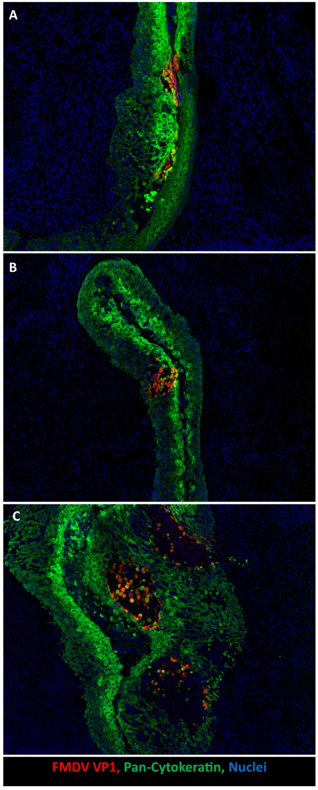
Figure 2.

Development of microvesicles within oropharyngeal tonsil epithelium during early infection. (A) Earliest detection of infection occurs within paraepiglottic tonsil at 24 h post intraoropharyngeal inoculation. FMDV antigen (red) in clusters of infected cytokeratin-positive (green) epithelial cells in superficial layers of crypt epithelium. (B) At 48 h post intraoropharyngeal inoculation, a single microvesicle is present within the tonsil of the soft palate. Focus of FMDV-infected (red) epithelial cells expanding through deeper layers of epithelium (green). (C) At 78 h post intraoropharyngeal inoculation, three distinct microvesicles are present within crypt epithelium of the tonsil of the soft palate. Sloughed FMDV VP1/cytokeratin double positive cells are present in vesicle lumen. 10× magnification.
