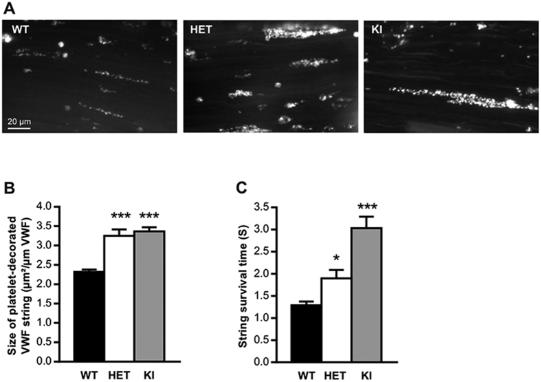
Figure 5. In vivo VWF/platelet string formation.
In vivo platelet-decorated VWF strings on the surface of mesenteric endothelium of WT-, HET- or KI-mice after topically application of FeCl3 solution (10%). (A) Images represent “platelet strings” attached to an injured vein. (B) The size of platelet-decorated VWF strings was calculated by dividing the length of each “platelet string” by their surface. (C) The lifetime of platelet-decorated VWF strings was determined per mouse and it was designated as “string survival time” (SST). In total, at least 200 platelet-decorated VWF strings were analyzed for each genotype. Statistical significance was determined by 1-way ANOVA followed by Dunnett’s test (*p < 0.05; ***p < 0.001). Scale bar represents 20 μm.

