
Figure 1. Crystal structure of p27/CDK2/CyclinA complex (PDB ID: 1JSU11).
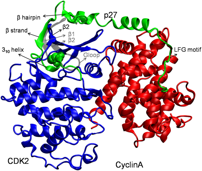
Color scheme is, CDK2 blue, Cyclin A red, and p27 green. Important regions of p27 are labeled and the location of Tyr88 in 310 helix of p27 is shown in sphere representation. The position of β2 strand of CDK2, which forms hybrid β-sheet with p27 β-strand is shown and labeled. Notably, the β1 strand and G-loop of CDK2 were missing in this structure. For comparison, the position of β2 strand and that of the G-loop and β1 strand in active CDK2 is shown (in grey, also labelled in grey) from the crystal structure of CDK2/CyclinA complex (PDB ID: 1QMZ28) by superposing the two crystal structures.