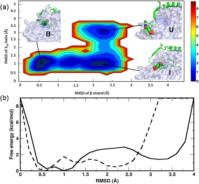
Figure 7. Free energy profile of the unbinding of phosphorylated p27.
(a) The unfolding free energy landscape of pY88-p27 from CDK2/CyclinA complex with associated color scale. Representative structures of the three major states are shown - the bound state (state-B), intermediate state (state-I), and unfolded state (state-U). (b) The 1D free-energy profiles of p27 unbinding based on Cα-RMSD of its β-strand (dashed lines) and 310 helix (solid line) as obtained from Boltzmann reweighting distribution.

