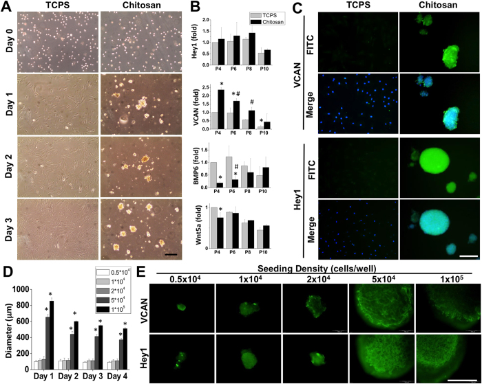
Figure 1.
Dermal papilla (DP, 1.3 × 10⁶ cells) cells were seeded on tissue culture polystyrene dish (TCPS) or chitosan-coated microenvironment (CCM) for 10-cm dish (Chitosan) to observe the sphere formation by acquiring the phase images at Day 0, 1, 2, and 3 (A). The adherent culture of DP cells caused the loss of DP characteristic, versican (VCAN), after multiple passages while the sphere formation maintained the DP markers, such as VCAN and Hey1 (B). The induction of VCAN and Hey1 protein expressions were confirmed by immunofluorescent staining by double staining with DAPI for cells seeded on TCPS and Chitosan coated dishes (C). Although the chitosan-coated dish maintained the DP characteristics, high variability of sphere size and protein expression patterns were observed in both VCAN and Hey1 staining. To control the sphere size, the CCM was created on the 96-well culture plate and seeding cell with different density for each well for measuring the DP sphere size at Day 1, 2, 3, and 4 (D). When the seeding density exceeded 5 × 10⁴ cells per well, the DP cells formed a single sphere in each well and the sphere condensed during culture. The immunofluorescent staining of VCAN and Hey1 indicated the sphere derived from a higher seeding density than 5 × 10⁴ cells promoted better DP characteristics (E). *In (B) significant difference from the adherent DP cells at passage 4, p < 0.05. #In (B) significant difference from the adherent DP cells at same passage, p < 0.05. *In (D) significant difference from seeding 0.5 × 10⁴ cells, p < 0.05. Scale bar = 200 μm.

