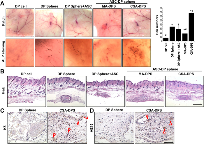
Figure 4. Hair induction was tested by in vivo patch assay in nude mice using suspending DP cells, DP spheres, DP spheres with suspending ASCs (DP Sphere + ASC), ASC and DP mixed spheres (MA-DPS), and the core-shell spheres with DP core and ASC shell (CSA-DPS).
The CSA-DPS showed a highly pigmented patch, high ALP staining in whole mount tissue, and the most vivid hair number after 4 weeks of injections (A). H&E staining showed the success of hair follicle neogenesis in hypodermis (B). Distributions of brown color in immunohistochemistry (IHC) staining indicated expression of K5 for the outer root sheath of HF (arrow heads) (C). To confirm the hair-like structure in CSA-DPS, the inner root sheath was also identified by AE15 staining (D). *Significant difference from suspending DP cells, p < 0.05. #Significant difference from DP sphere, p < 0.05. Scale bar in (B) = 500 μm. Scale bar in (B) = 100 μm.

