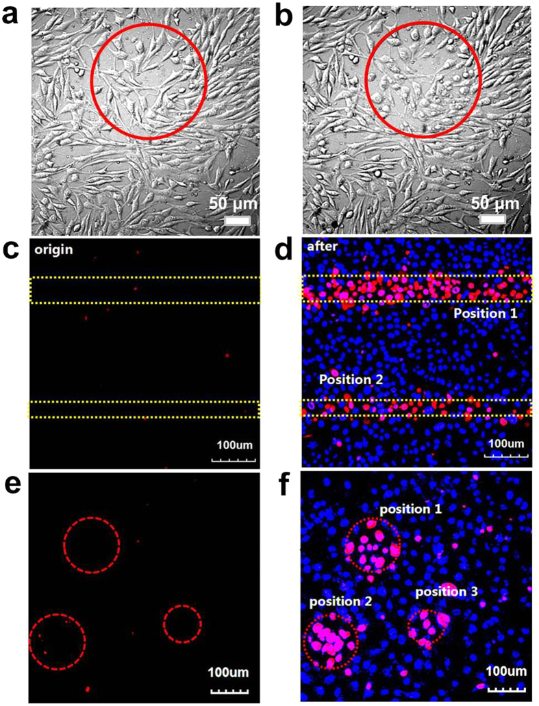
Figure 6. Laser-induced cell detachment.
Confocal micrograph of mouse fibroblast 3T3 cells growing on a (gold-collagen nanoconjugates/TPPAc-PLL)5 film (a), after irradiation with a laser (559 nm, 10 min, light intensity of 40%, 4.0 μs/Pixel) (b). Fluorescence micrographs of cells before (c,e) and after laser irradiation (559 nm, 10 min, light intensity of 40%, 4.0 μs/Pixel) (d,f ). The cells were stained with Hoechst 33342 (staining cell nuclei) and propidium iodide (PI, staining nuclei of dead cell). The regions marked by the red circles and yellow dashed lines are exposed to the laser irradiation.

