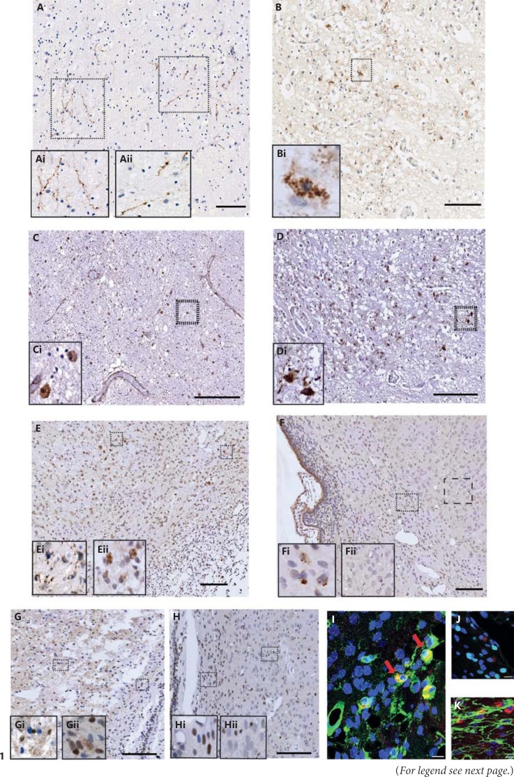
Fig. 1.
Cleaved caspases in the human preterm brain and in a neonatal hypoxic-ischaemic rat model. Immunoreactivity of cl-C6 (A, B) and cl-C3 (C, D) was expressed in a human preterm white matter injury case in the periventricular white matter (A, C) and anterior limb of the internal capsule (B, D). cl-C6 staining appeared fibrous (Ai, Aii) and perinuclear (Bi). Sporadic and nuclear cl-C3 was seen in the periventricular white matter region (C, Ci). However, the internal capsule the cl-C3 immunoreactivity was both nuclear and cytoplasmic (D). Following neonatal hypoxia-ischaemia in rats, cl-C6 (E, F) was observed in both the ipsilateral (E) and contralateral (F) hemispheres. Consistent with the observations made in the human tissue, cl-C6 again appeared to be fibrous in the cortex (Ei) and perinuclear in white matter (Eii) in the ipsilateral hemisphere, whereas in the contralateral side, there was perinuclear staining seen in the white matter (Fi) but not in the cortex (Fii). cl-C6 immunoreactivity was co-localised mainly in the microglia (tomato lectin; G) but not with oligodendroglia (Olig-2; H) or astroglia (GFAP; I). cl-C3 was also observed in rats following neonatal hypoxia-ischaemia, (J, K). Nuclear and punctate staining of cl-C3 was abundant in the ipsilateral side (J, Ji, Jii), but the contralateral side had primarily nuclear staining (K, Ki, Kii). Red arrows indicate co-labelled cells. Scale bar = 100 µm (A-F, J, K) and 10 µm (G-I).

