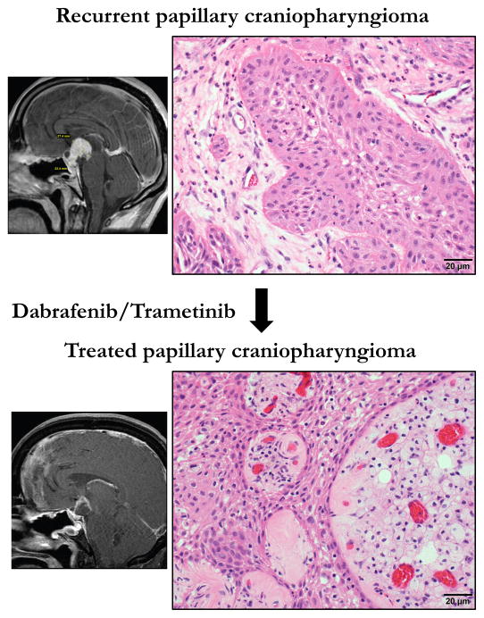
Figure 1.
H&E stained sections of pre- and post-treatment papillary craniopharyngioma from case reported in Brastianos et al., JNCI 2015 48. Top panel shows the recurrent tumor. The lower panel shows the tumor following treatment with dabrafenib and trametinib. Side panels are sketch renditions of MRI images from our exceptional responder patient 48.

